
Introduction: The Difference Between Digital Radiology and Traditional Radiographic Imaging
Before delving into specific types, it's essential to clarify the core differences between digital radiology systems and traditional film-based radiographic imaging. Traditional radiographic imaging relies on film processing, a time-consuming process involving harmful chemicals, and offers lower image resolution. Digital radiology systems, on the other hand, directly convert X-ray energy into digital signals. This digital revolution brings numerous advantages: higher image quality, lower radiation doses to patients, faster image acquisition and sharing, and seamless integration with electronic health records. As a result, digital X-ray equipment has become standard equipment in the vast majority of medical institutions, from small clinics to large hospitals.
In this article, we will explore the main types of digital radiology systems, focusing on their core principles and practical applications. We will also explain key terms such as digital X-ray systems, portable digital X-ray machines, and digital radiographic imaging X-ray machines to help you fully grasp this important medical technology.
Imaging technology is the fundamental criterion for categorizing digital radiology systems, as it determines the core working principle, image quality, and clinical suitability of each system. Each type of system developed based on different imaging technologies has its own unique advantages and limitations, making it tailored to specific diagnostic scenarios. Below, we analyze the four most common types of digital radiology systems by imaging technology, including their core principles and practical application scenarios, and explain how they integrate with x ray machine digital radiography and digital x ray equipment ecosystems.
Computed Radiography (CR) was the first commercially successful digital x-ray system that bridged the gap between traditional film radiography and fully digital radiology solutions, and it remains a vital part of digital x ray equipment in many healthcare facilities to this day. As a classic form of x ray machine digital radiography, CR revolutionized medical imaging by eliminating the need for chemical film processing while still being compatible with the existing x-ray equipment infrastructure of traditional radiology rooms, which made it a cost-effective digital transformation option for many medical institutions.
The core of the CR system is the photostimulable phosphor (PSP) plate, which replaces the traditional x-ray film as the image capture medium. When x-ray photons pass through the patient’s body and hit the PSP plate, the plate’s phosphor crystals absorb the x-ray energy and store it as a latent image—an invisible image formed by the excited electrons in the crystals. After the exposure, the PSP plate is inserted into a CR reader, where a high-precision laser beam scans the plate line by line. This laser stimulation causes the excited electrons to release the stored energy in the form of visible light photons. A photomultiplier tube (PMT) in the reader captures these light photons and converts them into electrical signals, which are then digitized, processed, and enhanced by a computer to generate a clear digital image. The PSP plate can be erased and reused after scanning, making the CR system more environmentally friendly and cost-efficient than film-based radiography.
CR systems are valued for their versatility and compatibility, making them ideal for healthcare facilities that are in the process of transitioning from traditional film to digital radiography without a full-scale replacement of existing equipment. They are widely used in primary care clinics, rural hospitals, and small medical centers for routine diagnostic imaging, including chest, abdominal, skeletal, and extremity radiography. CR is also a popular choice for low-to-medium volume imaging departments, as it balances performance and cost effectively. In addition, CR systems are often used in emergency departments for temporary imaging needs and in outpatient clinics for basic diagnostic checks, as they are easy to operate and require minimal training for radiology technicians. For medical facilities with limited budgets, CR serves as an accessible entry point to x ray machine digital radiography, allowing them to enjoy the benefits of digital imaging—such as electronic image storage and remote sharing—without the high upfront investment of more advanced digital systems.
Direct Digital Radiography (DR) is the most advanced and widely adopted digital x-ray system in modern healthcare, representing the gold standard of x ray machine digital radiography. As a flagship product of digital x ray equipment, DR has surpassed CR in almost all key performance indicators, offering faster image acquisition, higher resolution, lower radiation doses, and more intuitive operation. DR systems have become the primary choice for large hospitals, high-volume imaging centers, and tertiary medical institutions, driving the digital transformation of radiology departments worldwide.
Unlike CR, which uses a PSP plate and post-exposure scanning for digitization, DR systems feature an integrated flat-panel detector (FPD) that converts x-ray energy directly into digital electrical signals in real time, eliminating the need for a separate reader and post-processing steps. The flat-panel detector is composed of thousands of tiny pixel sensors, a thin-film transistor (TFT) array, and either a direct or indirect conversion layer. Indirect conversion DR detectors use a scintillator (e.g., cesium iodide or gadolinium oxysulfide) to convert x-ray photons into visible light, which is then detected by the TFT array and converted into electrical signals. Direct conversion DR detectors use an amorphous selenium layer that directly converts x-ray photons into electrical charges, which are then collected by the TFT array for digitization. In both cases, the electrical signals are immediately transmitted to a computer, which processes and displays the digital image on a monitor within 1-3 seconds—far faster than CR systems. This real-time imaging capability is the most distinctive advantage of DR, as it allows radiology technicians to check image quality instantly and retake images if needed, significantly improving workflow efficiency.
DR systems are suitable for almost all clinical imaging scenarios, from routine outpatient checks to complex inpatient diagnostics, and are widely used in chest, orthopedic, pediatric, neonatal, and dental radiography. Their high-resolution imaging and low radiation dose make them particularly ideal for pediatric radiology, where minimizing radiation exposure is a top priority. In large hospitals and trauma centers, DR systems are the backbone of emergency imaging, as they can quickly capture clear images of fractures, internal bleeding, and other traumatic injuries, enabling rapid diagnosis and treatment. DR is also used in specialized imaging departments, such as mammography, where dedicated digital mammography DR systems deliver high-contrast images for early breast cancer screening. Additionally, DR systems can be seamlessly integrated with Picture Archiving and Communication Systems (PACS) and electronic health record (EHR) systems, allowing digital images to be stored, shared, and accessed remotely by healthcare providers across different departments or even different medical facilities. As the core of digital x ray equipment, DR systems have become an essential investment for healthcare facilities aiming to improve diagnostic accuracy, streamline workflow, and enhance patient care.
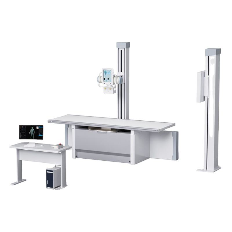
ArKang provides durable digital x-ray systems with outstanding high-resolution imaging performance. The AKX500D is one of our premium digital x-ray equipment models, supporting comprehensive full-body imaging for the chest, abdomen, limbs and other anatomical parts, while also offering a highly cost-effective price.
Digital Fluoroscopy (DF) is a specialized digital x-ray system designed for dynamic real-time imaging, a key difference from the static imaging provided by CR and DR systems. As a unique type of digital x ray equipment, DF enables radiologists to observe the movement of internal organs, blood flow, and the passage of contrast media in the body in real time, making it an indispensable tool for interventional radiology and functional diagnostic imaging.
The core principle of DF systems is continuous x-ray exposure and real-time digital image capture and display. A DF system consists of an x-ray tube, an image intensifier or flat-panel detector, a camera system, and a digital processing unit. The x-ray tube emits a low-dose, continuous x-ray beam that passes through the patient’s body and hits the detector. The detector converts the x-ray energy into electrical signals, which are then digitized and processed by the computer to generate a continuous stream of digital images—essentially a “moving picture” of the internal body structures. Early DF systems used image intensifiers and analog cameras, but modern DF systems have adopted flat-panel detectors (FPD-DF), which deliver higher resolution, lower radiation doses, and better image quality than traditional image intensifier-based systems. The digital images are displayed on a high-definition monitor in real time, and radiologists can record, pause, and replay the image sequence for detailed analysis. DF systems also support the injection of contrast media to enhance the visibility of specific organs or blood vessels, further expanding their diagnostic capabilities.
DF systems are primarily used for functional and interventional diagnostic imaging, where real-time observation of internal body movement is required. The most common applications include gastrointestinal (GI) imaging, such as barium swallows, barium enemas, and upper GI series, where radiologists can observe the peristalsis of the esophagus, stomach, and intestines and detect abnormalities like ulcers, strictures, or tumors. DF is also widely used in interventional radiology procedures, such as catheter insertions, angiography, and minimally invasive surgical guidance, where radiologists need real-time imaging to navigate medical instruments through the body. In orthopedics, DF is used to guide fracture reduction and joint injections, ensuring accurate placement of implants and needles. Additionally, DF is used in urology for intravenous pyelography (IVP) to observe the flow of urine from the kidneys to the bladder, and in cardiology for basic cardiac imaging to assess heart function. As a critical part of digital x-ray systems, DF fills the gap of static imaging and provides radiologists with a dynamic, real-time view of the body, making it essential for a wide range of specialized clinical procedures.

For example, ArKang’s AK-RF65D is a high-performance Digital X-Ray System with Dynamic Fluoroscopy that supports real-time, continuous imaging. In addition, ArKang backs this system with a one-year warranty and comprehensive after-sales service.
Digital Subtraction Angiography (DSA) is a highly specialized branch of digital fluoroscopy and a sophisticated digital x-ray system dedicated to vascular imaging. As a premium digital x ray equipment, DSA is designed to eliminate the interference of bones, soft tissues, and other non-vascular structures from the image, leaving only the blood vessels in high contrast and detail. This unique capability makes DSA the gold standard for diagnosing and treating vascular diseases, and it is a core tool in cardiology, neurology, and vascular surgery departments.
The core principle of DSA is the “subtraction” technique, which involves capturing two sets of digital images and subtracting one from the other to isolate the blood vessels. The first set of images, called the “mask image,” is captured before the injection of iodinated contrast media into the patient’s bloodstream—this image shows all the body structures (bones, soft tissues, and blood vessels) in the imaging area. The second set of images, called the “contrast image,” is captured immediately after the contrast media is injected, as the media flows through the blood vessels and makes them visible against the surrounding tissues. The DSA system’s computer then subtracts the mask image from the contrast image, pixel by pixel, which removes all the non-vascular structures that appear in both images. The result is a high-contrast digital image that shows only the blood vessels, with no interference from bones or soft tissues. Modern DSA systems also feature 3D reconstruction capabilities, which can generate three-dimensional vascular images from multiple 2D subtraction images, providing radiologists with a more comprehensive view of the vascular anatomy.
DSA systems are exclusively used for vascular imaging and interventional vascular procedures, and their applications span cardiology, neurointerventional radiology, and peripheral vascular surgery. In cardiology, DSA is the gold standard for coronary angiography, which is used to diagnose coronary artery disease by visualizing blockages, narrowings, or aneurysms in the coronary arteries that supply blood to the heart. DSA is also used to guide interventional cardiac procedures, such as angioplasty and stenting, where radiologists use real-time vascular images to navigate catheters and place stents to open blocked arteries. In neurointerventional radiology, DSA is used to image the cerebral blood vessels, diagnosing conditions like cerebral aneurysms, arteriovenous malformations (AVMs), and ischemic strokes, and guiding minimally invasive treatments such as coil embolization for aneurysms. In peripheral vascular surgery, DSA is used to image the blood vessels in the arms, legs, abdomen, and kidneys, detecting peripheral artery disease (PAD) and guiding interventional procedures to treat blocked or narrowed peripheral vessels. As a highly specialized digital x-ray system, DSA is a critical investment for tertiary hospitals and specialized medical centers, enabling accurate diagnosis and minimally invasive treatment of vascular diseases and significantly improving patient outcomes.
In addition to imaging technology, mobility is another important criterion for classifying digital radiology systems and digital x ray equipment. The mobility of a system determines where it can be used—whether it is permanently installed in a dedicated radiology room or can be moved to the patient’s bedside, emergency scenes, or remote medical facilities. This classification is particularly important for healthcare facilities that need to provide imaging services to bedridden, critically ill, or immobile patients, as well as for mobile medical services in remote areas. Below, we analyze three types of digital radiology systems by mobility: fixed systems, the portable digital x ray machine, and special mobility systems, including their advantages and application scenarios.
Fixed digital radiology systems are the most common type of digital x-ray system in healthcare facilities, and they are the main component of x ray machine digital radiography in dedicated radiology rooms. These systems are permanently installed and fixed to the floor, walls, or ceiling of a specially designed radiology room, with a dedicated x-ray tube, detector, and imaging table. Fixed systems are built for high performance, high-volume imaging, and consistent image quality, making them the backbone of radiology departments in hospitals and imaging centers.
The primary advantage of fixed digital radiology systems is their superior performance and image quality. They are equipped with high-power x-ray generators, large-area high-resolution flat-panel detectors, and advanced image processing software, which deliver sharp, high-contrast images even for complex imaging scenarios, such as chest and abdominal radiography for obese patients. Fixed systems also offer greater stability and consistency than mobile systems, as they are not subject to movement or vibration, ensuring that every image is of the same high quality. Additionally, fixed digital radiology systems can be fully integrated with other digital x ray equipment and hospital information systems, including PACS, EHR, and radiology information systems (RIS), streamlining the entire imaging workflow from image capture to diagnosis and report generation. Another key advantage is their customizable design—fixed systems can be equipped with additional features such as automatic patient positioning, dose reduction technology, and 3D reconstruction, tailoring to the specific needs of different imaging departments. Finally, fixed systems have a longer service life and lower maintenance costs than mobile systems, as they are not exposed to the wear and tear of frequent movement.
Fixed digital radiology systems are designed for high-volume, routine, and complex diagnostic imaging in dedicated radiology rooms, and they are widely used in large hospitals, tertiary medical centers, and professional imaging centers. They are the primary choice for all types of static imaging (CR/DR) and dynamic imaging (DF/DSA), including chest, abdominal, skeletal, pediatric, and mammography radiography, as well as interventional radiology procedures. Fixed DR systems are used in general radiology departments for routine outpatient and inpatient imaging, while fixed DSA systems are installed in dedicated interventional radiology suites for vascular imaging and minimally invasive procedures. Fixed digital radiology systems are also used in specialized imaging departments, such as dental radiology and orthopedic radiology, where dedicated fixed systems deliver high-resolution images for specialized diagnostic needs. For healthcare facilities with a high volume of imaging patients, fixed systems are the most efficient and cost-effective option, as they can handle a large number of patients per hour and deliver consistent, high-quality images for accurate diagnosis.
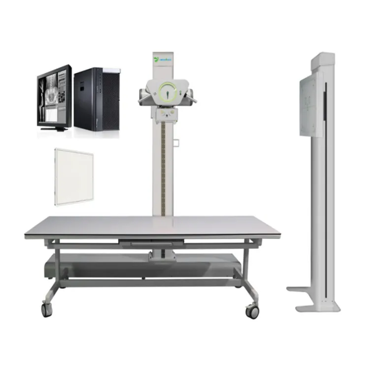
Fixed digital x-ray systems typically adopt a dual-column structure, delivering excellent stability and enhanced performance. ArKang offers static digital x-ray machines with a full range of power options to meet all kinds of clinical needs.
The portable digital x ray machine is a compact, mobile digital x-ray system and a highly versatile piece of digital x ray equipment that has transformed bedside and point-of-care imaging. Unlike fixed systems, the portable digital x ray machine is lightweight, battery-powered, and equipped with wheels, allowing radiology technicians to move it directly to the patient’s bedside, emergency scenes, or remote medical facilities—eliminating the need to transport immobile, critically ill, or injured patients to the radiology room. In recent years, advances in technology have made modern portable digital x ray machine models comparable to fixed systems in terms of image quality, while still maintaining their compact and mobile design, making them an essential tool in modern healthcare.
The most significant advantage of the portable digital x ray machine is its unparalleled mobility and flexibility. It can be used in any clinical setting, including intensive care units (ICUs), neonatal intensive care units (NICUs), general wards, operating rooms, emergency departments, and even outside the hospital (e.g., ambulance, disaster relief scenes, and remote rural clinics). This mobility eliminates the risk and discomfort of transporting critically ill, bedridden, or post-surgical patients to the radiology room, which is a major benefit for both patients and healthcare providers. Modern portable digital x ray machine models also deliver high-quality digital images that are comparable to fixed DR systems, thanks to advanced flat-panel detectors and low-dose imaging technology. They are also easy to operate, with intuitive controls and a lightweight design that requires only one or two technicians to move and operate. Additionally, the portable digital x ray machine is equipped with wireless connectivity, allowing digital images to be transmitted to PACS or EHR systems in real time, enabling radiologists to diagnose images remotely and provide timely treatment recommendations. Another advantage is its cost-effectiveness—portable digital x ray machine models have a lower upfront investment than fixed systems, making them an accessible option for small clinics, nursing homes, and mobile medical services.
The portable digital x ray machine is primarily used for bedside imaging and point-of-care diagnostic checks, making it an essential tool in ICUs, NICUs, and general wards for monitoring critically ill patients—such as those with respiratory failure, heart disease, or severe trauma—by capturing chest x-rays and other images without moving the patient. It is also widely used in emergency departments and ambulances for on-site imaging of trauma patients, such as those with fractures, chest injuries, or head trauma, enabling emergency physicians to make rapid diagnostic decisions and initiate treatment on the spot. In operating rooms, the portable digital x ray machine is used for intra-operative imaging to guide surgical procedures, such as fracture fixation and implant placement. Beyond hospital settings, the portable digital x ray machine is used in nursing homes, home healthcare, and mobile medical clinics to provide imaging services to elderly, immobile, or homebound patients. It is also a critical tool in disaster relief and remote medical missions, where access to a dedicated radiology room is unavailable. For rural hospitals and small clinics with limited space, the portable digital x ray machine serves as a multi-purpose digital x ray equipment that can handle all routine imaging needs, eliminating the need for a dedicated radiology room. As a key part of digital x-ray systems, the portable digital x ray machine has greatly expanded the reach of medical imaging, ensuring that high-quality diagnostic services are available to patients wherever they are.
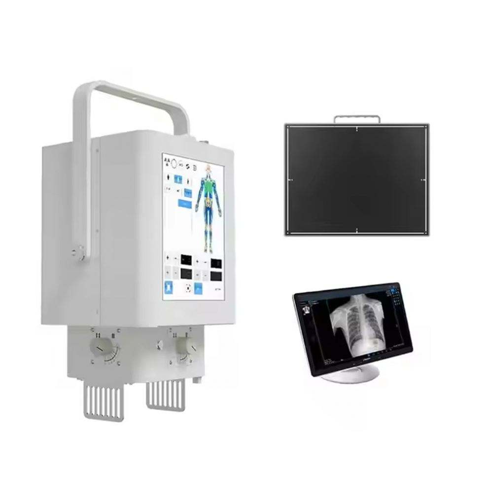
The price of the portable digital x ray machine varies significantly depending on multiple factors. At ArKang, you can get the most cost-effective portable digital x ray machine to meet your clinical needs.
In addition to fixed systems and the portable digital x ray machine, there are a range of special types of digital radiology systems that combine unique imaging technologies and mobility features, designed to meet the specialized diagnostic needs of specific clinical scenarios. These special systems are advanced digital x ray equipment that integrate the advantages of fixed and mobile systems, or feature unique imaging capabilities that standard systems do not have, making them ideal for specialized radiology departments and clinical procedures.
The primary advantage of special digital radiology systems is their specialized functionality, which is tailored to specific diagnostic and clinical needs that standard fixed or portable digital x-ray systems cannot meet. For example, dual-energy digital radiology systems use two different x-ray energies to capture images, allowing radiologists to distinguish between different types of tissues (e.g., bone, soft tissue, and calcifications) and detect abnormalities that are not visible on standard x-rays. Cone Beam Computed Tomography (CBCT) systems, a special type of digital radiology system, generate 3D volumetric images with high spatial resolution, which is far more detailed than the 2D images provided by standard CR/DR systems. Another advantage is their hybrid mobility—some special systems are semi-portable, meaning they are compact enough to be moved between different rooms in a hospital but still deliver the high performance of fixed systems. Additionally, special digital radiology systems often feature the latest dose reduction technology and AI-powered image processing, which deliver high-quality images with even lower radiation doses and enable automated image analysis and abnormality detection. Finally, these systems can be integrated with other advanced medical equipment, such as surgical navigation systems and robotic surgery systems, making them essential for minimally invasive and precision medicine procedures.
Special digital radiology systems are used in specialized clinical and diagnostic scenarios across different medical fields. CBCT systems are widely used in dental radiology, orthodontics, and maxillofacial surgery, where 3D images are essential for dental implant placement, orthodontic treatment planning, and facial fracture repair. They are also used in orthopedic surgery for 3D imaging of bones and joints, enabling precise surgical planning and implant placement. Dual-energy digital radiology systems are used in chest radiography to detect lung nodules, in urology to detect kidney stones, and in oncology to distinguish between benign and malignant soft tissue tumors. Semi-portable DF systems are used in operating rooms for intra-operative dynamic imaging, guiding minimally invasive surgical procedures such as laparoscopic surgery and robotic surgery. Photon-counting digital radiology systems, a cutting-edge special type of digital x-ray system, are used in specialized imaging centers for high-resolution imaging of small structures (e.g., microvasculature and small lung nodules), enabling early detection of diseases that are not visible on standard digital radiology systems. These special systems are also used in pediatric radiology and neonatal radiology, where their low-dose imaging technology and high-resolution capabilities are ideal for minimizing radiation exposure in children and capturing clear images of small, delicate body structures. As the field of medical imaging continues to advance, special digital radiology systems are becoming an increasingly important part of digital x ray equipment, driving the development of precision medicine and minimally invasive surgery.
Digital radiography systems have redefined modern medical imaging, replacing traditional film-based radiography. With their high resolution, low radiation dose, rapid imaging speed, and digital workflow, they have become an indispensable part of healthcare. From the classic CR systems that pioneered digital imaging to the advanced DR, DF, and DSA systems that set current clinical standards, and from high-performance stationary systems to multifunctional portable digital X-ray machines, each digital X-ray system has its unique core principles, advantages, and application scenarios, designed to meet the diverse diagnostic needs of clinical practice.
Follow Us


