Introduction
Every X-ray image tells two stories. The first is the clinical story — the bone, the lung, the dental root that a radiologist needs to see clearly. The second is a quieter story about the machine that produced it: how much power it could deliver, how precisely it could focus the beam, and how much stray radiation it scattered into the room around the patient. For anyone buying an X-ray machine — a veterinary clinic upgrading its imaging, a hospital outfitting a bedside unit, an orthopedics practice opening a new wing, or a distributor building a product lineup — these two stories decide whether the investment is wise.
X-ray machines come in three broad categories: portable, mobile, and fixed. They share the same fundamental physics — an X-ray tube emits photons that pass through tissue and expose a detector — but they differ enormously in power, image quality, scatter radiation behavior, and the settings where each one truly earns its keep. This article compares the three types head to head, focusing on the two factors that matter most in practice: image quality and scatter radiation. Along the way we will look at how modern trends — digital detectors, AI-assisted enhancement, low-dose technology, and remote reading — are reshaping what each machine can deliver.
Whether you are a first-time buyer, a seasoned radiologic technologist, or a seller trying to match a product to a customer, this guide gives you a structured way to compare. By the end you will know which machine type fits which job, what hidden costs to expect, and how to protect patients and staff from scatter radiation no matter which unit you choose.
1.The Three Types at a Glance
Before comparing image quality and scatter radiation, we need a clear picture of what each machine type actually is.
Portable X-ray machines are the lightweights. A true portable unit — often handheld or mounted on a small cart — is designed to move freely between rooms, and sometimes out into the field, a barn, or a disaster site. Portable machines typically operate at low to moderate power, with tube voltages in the 40–100 kVp range and relatively modest milliamperage. They use small, lightweight tubes and usually connect to a flat-panel digital detector. Their strength is flexibility: they go where the patient is. Their tradeoff is that they cannot match the raw power or image quality of a room-based system, and they place the operator close to the X-ray field.
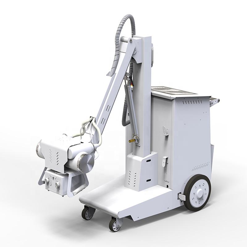
Mobile X-ray machines are the middle ground. These are wheeled, self-contained units — the classic "portable in the hospital" cart — that radiology departments wheel into patient rooms, intensive care units, emergency departments, and operating suites. Mobile units are more powerful than their truly portable cousins, typically delivering 30–50 kW with a wider kVp range, and they carry their own generator and often a dedicated DR panel. They are the workhorses of bedside imaging: when a ventilator-bound ICU patient cannot be moved to the radiology suite, the mobile unit comes to them.

Fixed X-ray machines are the flagship. Installed permanently in a dedicated radiology room, fixed units are ceiling- or floor-mounted and connected to a high-capacity generator, usually 50–80 kW or more. They offer the widest kVp and mAs ranges, the most powerful tubes, the most precise collimation, and the best access to image-sharpening accessories such as anti-scatter grids. Because the room can be designed around the machine, fixed systems pair with lead-lined walls, remote control stations, and built-in shielding that no portable or mobile unit can enjoy.
|
Feature
|
Portable
|
Mobile
|
Fixed
|
|
Typical power
|
Low (a few kW)
|
Moderate (30–50 kW)
|
High (50–80+ kW)
|
|
Tube voltage range
|
Narrow (40–100 kVp)
|
Medium
|
Wide (up to 150 kVp)
|
|
Placement
|
Handheld / small cart
|
Wheeled cart
|
Ceiling- or floor-mounted
|
|
Dedicated room needed
|
No
|
No
|
Yes
|
|
Typical settings
|
Field, home, small vet clinic
|
ICU, ER, OR, bedside
|
Radiology department
|
|
Grid access
|
Limited
|
Possible
|
Standard
|
|
Operator distance
|
Very close
|
Close
|
Remote control room
|
With these definitions in place, we can turn to the two questions that most buyers care about: how good is the image, and how much scatter radiation does the machine produce?
2.X-Ray Image Quality: How Each Machine Type Performs
Image quality is the reason anyone buys an X-ray machine in the first place. A radiograph is only useful if the radiologist can distinguish subtle fractures, early lesions, or the fine structure of soft tissue. Understanding why the three machine types differ begins with the six factors that govern image quality:
-
Tube voltage (kVp) — higher voltage gives the beam more penetration and can improve contrast in dense anatomy, but too high a voltage washes out low-contrast details.
-
Tube current–time product (mAs) — more mAs means more photons and less quantum noise, but also longer exposure time.
-
Focal spot size — a smaller focal spot produces sharper images because it reduces geometric unsharpness.
-
Detector type — modern flat-panel digital detectors (DR) capture far more detail than older computed radiography (CR) systems, and detector resolution directly sets the ceiling on image detail.
-
Source-to-image distance (SID) — greater distance improves geometric accuracy and reduces magnification distortion.
-
Anti-scatter grid — a grid absorbs scattered photons before they hit the detector, sharpening contrast at the cost of some dose.
Now let us see how the three machine types perform against these factors.
2.1 Portable X-Ray Image Quality
Portable machines make real, practical compromises to achieve their size. Because the entire unit must be lightweight, its tube is small, its generator is modest, and its power output is limited. The practical consequences are straightforward.
First, low power forces longer exposure times. To accumulate enough photons for a readable image, the machine must keep the tube energized longer — and any patient movement, breathing, or tremor during that window creates motion blur. This is why portable chest or abdominal images in a moving or uncooperative subject can come out soft.
Second, small focal spots and limited kVp ranges constrain contrast. Portable units cannot always dial in the ideal tube voltage for dense anatomy such as an adult pelvis or a large veterinary patient. The operator often must accept a compromise between penetration and contrast that a larger machine would not require.
Third, portable units rarely accommodate anti-scatter grids well. Without a grid, scattered radiation reaches the detector and lowers contrast, which further erodes image quality in thicker body parts.
None of this means portable images are bad — far from it. Modern portable DR units with good flat-panel detectors produce perfectly adequate images for screening, extremity work, dental, and small-to-medium veterinary patients. The point is that the ceiling is lower. A portable machine can do a great job within its comfort zone, but it cannot match the consistency of a fixed system on large, dense, or motion-prone anatomy.
2.2 Mobile X-Ray Image Quality
Mobile units sit between portable and fixed, and so does their image quality. Because a cart can carry a heavier generator than a handheld device, mobile machines typically deliver significantly more mAs and a wider kVp range. This means shorter exposure times, less motion blur, and more consistent penetration of dense anatomy than portable units achieve.
The quality of mobile imaging hinges on the detector. Most mainstream mobile X-ray systems are now equipped with high-performance digital radiography (DR) flat-panel detectors. For instance, when a mobile X-ray unit incorporates a high-quality detector—such as the Arkang Mars1717V wireless flat-panel detector—for bedside chest radiography, the diagnostic quality of the resulting images becomes comparable to that of fixed, room-based systems. This is precisely why mobile units are the standard choice for bedside imaging in ICUs and emergency departments, where the ability to reliably identify subtle clinical signs—such as changes in pulmonary ventilation or the precise positioning of medical tubes and lines—is essential.
The remaining gap between mobile and fixed is usually grid access and geometry. Some mobile units can mount anti-scatter grids, but many bedside exams are done without them, accepting slightly lower contrast in exchange for speed and simplicity. And because the machine must be positioned manually beside the bed, source-to-image distance and centering are less repeatable than in a dedicated room — so image quality depends somewhat on operator skill.
2.3 Fixed X-Ray Image Quality
Fixed machines represent the diagnostic gold standard, and the reasons are concrete rather than vague.
High generator power means high mAs with short exposure times, which freezes motion and delivers a photon-rich, low-noise image. A wide kVp range lets the technologist fine-tune penetration for everything from a pediatric hand to a heavy-torso chest. The fixed room provides repeatable, calibrated geometry: consistent source-to-image distance, precise centering, and a stable patient-table alignment that removes a major source of operator-dependent variability.
Most important, fixed rooms are where anti-scatter grids are used routinely and effectively. By absorbing scattered photons before they reach the detector, the grid dramatically improves contrast in large body parts — and that contrast is exactly what a radiologist needs to spot subtle pathology. When all these factors combine, a fixed-room image is the benchmark against which portable and mobile images are measured.
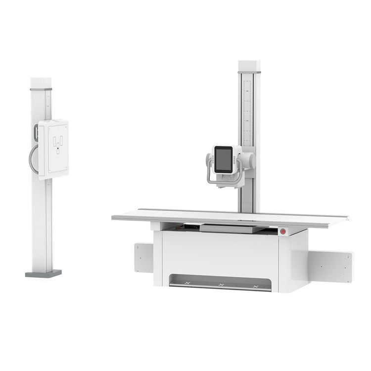
The AKX-iDR65 is a best-selling stationary X-ray system from ArKang. With a maximum power of 65kW, it supports comprehensive radiographic examinations of various body parts, including the chest, abdomen, spine, and extremities. The system features a laterally movable examination table and a vertical stand; the X-ray tube is rotatable, enabling multi-angle imaging and accommodating a wide range of patient positioning requirements, with the capability to detect even minute lesions. Offering an excellent price-to-performance ratio, this model is highly well-suited to the procurement needs of markets in Africa and Southeast Asia.
The AI wildcard. A genuinely hot trend is changing this equation: AI-assisted image enhancement. Modern software can denoise low-exposure images, sharpen edges, and reduce artifacts that used to require a powerful generator to fix. Some portable and mobile systems now ship with AI post-processing that narrows the quality gap with fixed machines. The implication matters to buyers: the image-quality disadvantage of a portable unit is shrinking, though it is not gone — physics still favors the bigger machine for dense anatomy.
3.Scatter Radiation: The Hidden Risk in Every X-Ray Exam
Image quality is only half the story. The other half is scatter radiation — the stray X-ray photons that are deflected as the primary beam passes through the patient. Understanding scatter radiation is essential because it affects two things at once: it degrades image quality, and it is the principal radiation hazard to everyone in the room.
3.1 What Is Scatter Radiation?
When an X-ray beam strikes the body, most photons pass through or are absorbed. But a significant fraction are deflected — a process called Compton scattering — and travel in random directions around the patient. These scattered photons carry energy outward into the room. They do two kinds of damage. First, if they reach the image detector, they fog the image and wash out contrast — the more scatter, the grayer the image. Second, they expose everyone standing nearby: the technologist, the nurse, the veterinarian holding an animal, or the family member assisting.
The amount of scatter depends mainly on three things: the thickness and size of the body part being imaged, the size of the irradiated field (collimation), and the beam energy. Larger patients, larger fields, and certain energy levels all generate more scatter. This is why imaging a heavy chest or an abdomen produces more scatter than imaging a hand.
3.2 How Each Machine Type Manages Scatter
Here the differences between portable, mobile, and fixed are sharp — and they are a major safety consideration.
Portable machines manage scatter the least well. By definition they are used outside a shielded room — in a home, a barn, an operating theater, or a clinic corridor. The operator stands close to the patient, often within the scatter field, with no lead-lined walls between them and the radiation. The machine's limited power and grid access mean more of what reaches the detector is scatter, and because the operator is near, personal protection becomes critical: lead aprons, thyroid collars, gloves, and distance are the only shields available. Field users must be especially disciplined about collimation and about positioning themselves outside the primary beam and as far from the patient as practical.
Mobile machines fall in the middle. They too are used at the bedside, so they lack a shielded room. However, mobile units often support better collimation and can accept grids, which reduce the scatter that reaches the detector. The operator still stands close, so the same PPE and distance discipline apply. The key difference is that mobile imaging is typically done in controlled hospital settings where technologists are trained in radiation safety, and where the scatter volume is somewhat lower because grids and collimation are more available.
Fixed machines manage scatter the best. The dedicated room is designed around the physics. Lead-lined walls absorb scatter before it reaches anyone. A remote control station lets the technologist stand behind shielding while making the exposure. Anti-scatter grids absorb scattered photons at the detector, simultaneously improving contrast and reducing the radiation burden on the image. Precise collimation restricts the beam to the area of interest, shrinking the scatter field. For the highest-examined and highest-exposure procedures, the fixed room is the gold standard of scatter control.
3.3 Scatter Radiation Levels: A Practical Comparison
It is tempting to think in terms of a single "scatter number" for each machine, but the honest answer is more nuanced: scatter depends far more on the patient, the collimation, and the exam than on the machine itself. A poorly collimated portable exam can scatter more than a well-run fixed-room study, and a thin patient scatters far less than a heavy one under any machine.
What is reliable to say is this: the machinery and environment for controlling scatter are best in a fixed room, middling on a mobile cart, and most demanding with a portable unit. The practical ranking of scatter control is therefore:
|
Machine type
|
Scatter control by design
|
Operator shielding
|
Typical exposure burden
|
|
Portable
|
Lowest (limited grid, no room)
|
PPE only, standing close
|
Highest per exam for operator
|
|
Mobile
|
Moderate (grid possible, hospital setting)
|
PPE, standing close
|
Moderate
|
|
Fixed
|
Highest (grid, collimator, lead room, remote station)
|
Remote control behind shielding
|
Lowest
|
The important principle — and a genuinely hot topic in radiation safety — is ALARA, which stands for "As Low As Reasonably Achievable." ALARA is not just a slogan; it is the guiding doctrine of X-ray safety. It means every imaging department should reduce radiation dose to patients and staff as far as practical, using distance, shielding, collimation, and the shortest necessary exposure. Buyers should ask themselves: can my staff practice ALARA with the machine I am buying? For a portable unit in the field, the answer demands extra care; for a fixed room, much of the work is done by the design.
4.Veterinary, Bedside, and Radiology: Matching the Machine to the Job
The right machine is the one that fits the job. Here is a practical match-up across the most common settings.
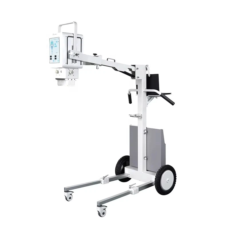
Veterinary imaging is one of the fastest-growing markets for X-ray, and it is dominated by portable and mobile units. Small-animal clinics love portables because they are affordable, mobile, and adequate for dogs and cats. Larger-animal practice — horses, cattle — often needs portable or mobile DR that can be moved to the animal rather than the reverse, since sedating and transporting a large animal is difficult and risky. The Arkang AK056-PE VET portable X-ray machine is a highly popular and representative model in the field of veterinary imaging. It is versatile enough for use on small to medium-sized companion animals—such as cats and dogs—while also being portable enough to be taken to farms for the examination of large livestock, including horses and cattle. A client in the Maldives purchased this unit for clinical diagnostic work on horses; following their initial use, they highly praised both its exceptional image quality and its intuitive, easy-to-master operation.
Bedside and intensive care is the home of the mobile unit. Patients on ventilators, in isolation, or too unstable to move need chest X-rays at the bedside, and the mobile cart delivers them with acceptable quality and manageable scatter when staff follow safety protocols.For example, Arkang’s AKX-mDR32 is a classic piece of mobile digital radiography equipment.
Emergency and orthopedic extremities are served well by portables. Fracture checks, dental X-rays, and quick screening exams in ERs, sports clinics, and dental practices are portable strengths.
The full radiology department is the domain of the fixed machine. High-volume, high-exposure, and high-quality-demanding work — chest, spine, abdomen, complex orthopedic studies — belongs in the dedicated room where image quality is highest and scatter is best controlled.
5. What's Trending in X-Ray Technology Right Now
A few developments are genuinely reshaping how buyers choose between portable, mobile, and fixed machines. If you are researching X-ray systems in 2025 and beyond, these are the conversations you will hear.
AI-assisted image enhancement. As noted, AI denoising and edge-sharpening can lift the quality of low-dose and portable images closer to fixed-room standards. This matters because it lets smaller machines produce cleaner images at lower radiation doses — a win for both image quality and scatter reduction.
Low-dose and dose-reduction technology. Manufacturers are competing on dose efficiency: automatic exposure control, better detectors that capture more of the beam, and software that extracts more signal from fewer photons. Low-dose capability is now a marketing differentiator across all three categories and directly supports the ALARA principle.
Wireless DR and portability. The shift from CR cassettes to flat-panel DR — and, more recently, to wireless, battery-powered panels — has made both portable and mobile units lighter, faster, and more practical in the field and at the bedside. Image quality has risen even as the hardware has shrunk.
Tele-radiology and remote reading. Images captured on any of these machines can now be transmitted to radiologists anywhere in the world. This elevates the importance of consistent, portable-friendly image formats and reliable network connectivity — and it means even a small clinic with a portable unit can access specialist interpretation.
Regulatory alignment. Buyers are increasingly checking compliance with standards such as IEC 60601 (general safety and essential performance of medical electrical equipment), and with FDA clearance (United States) or CE marking (European Union). Compliance is no longer a footnote; it is a purchase criterion, especially for cross-border buyers.
6.X-Ray Machine Cost: More Than the Price Tag
The purchase price is only the beginning. A complete comparison must account for what each machine type costs to own and operate.
Initial cost. Generally, portable machines are the least expensive, mobile units sit in the middle, and fixed systems are the most costly — sometimes by a wide margin.
Installation and room cost. This is where the gap widens most. A portable or mobile unit needs almost no infrastructure. A fixed machine requires a dedicated room, lead shielding, structural reinforcement for ceiling or floor mounting, power supply, and often regulatory inspection and licensing. For many buyers, this hidden cost — not the sticker price — is what decides the choice.
Maintenance and service. Fixed systems, with their larger generators and complex installations, may carry higher service costs, though portable units are not immune to repairs and calibration needs. Battery life is an ongoing cost for portable and mobile DR panels.
Training. Portable and mobile machines demand more operator judgment in positioning and radiation safety, so training costs can be higher per exam. Fixed rooms, with their repeatable geometry, make consistent technique easier.
Footprint and flexibility. A portable unit that serves three rooms may cost more in aggregate labor but far less in space than a fixed room serving one. For low-volume or multi-site practices, flexibility can outweigh the per-exam efficiency of a fixed system.
The takeaway: cost is a lifetime picture, not a sticker price. A buyer comparing "price" without counting the shielded room, maintenance, and training will underestimate the true cost of a fixed system — and overestimate the savings of a portable one.
7.How to Choose the Right X-Ray Machine
Choosing among portable, mobile, and fixed comes down to a small set of questions. Work through them in order.
-
What anatomy, and how large are the patients? Small extremities and small animals → portable is fine. Chest, spine, abdomen, and large patients at volume → you want at least mobile, ideally fixed.
-
Where is the exam performed? If patients cannot be moved (ICU, field, barn, home), you need portable or mobile. If you can build a dedicated room, fixed is the quality and safety choice.
-
What is the exam volume? Low volume and multi-location → portable or mobile to avoid idle capital. High volume in one location → fixed, for throughput and consistency.
-
What is your real budget, including the room and training? Count the shielded room, installation, maintenance, and training — not just the machine price.
-
Can your staff practice ALARA? If the machine will be used close to people without a shielded room, invest in PPE, training, and collimation discipline. If you can afford a remote-control fixed room, the safety burden drops dramatically.
-
What compliance does your market require? Check FDA, CE, IEC 60601, and local licensing before purchase, especially if you buy across borders.
As a rough rule of thumb: choose portable for mobility, low cost, small anatomy, and multi-site flexibility; choose mobile for bedside work in hospitals and medium-size patients; choose fixed for high-volume, high-quality, high-exposure diagnostic work where you can build a dedicated room.
8.Frequently Asked Questions
8.1 Is portable X-ray image quality as good as fixed X-ray image quality?
Generally, no. Fixed machines offer higher power, better grid access, and more repeatable geometry, so they produce higher contrast and resolution on large or dense anatomy. However, modern portable DR with AI enhancement can approach fixed quality on small-to-medium patients and extremities. Choose based on your anatomy mix.
8.2 How much scatter radiation does a mobile X-ray machine produce?
Scatter depends on patient size, field size, and beam energy rather than the machine alone. Mobile units used at the bedside do produce scatter to nearby staff, so operators should use lead aprons, thyroid collars, collimation, and distance. In hospital settings this is managed by trained technologists.
8.3 Can I use a portable X-ray machine for veterinary patients?
Yes. Portable DR units are widely used in small-animal practice and for large animals that cannot be moved. They are adequate for dogs, cats, and many equine and bovine studies, though very large patients may need a more powerful mobile or fixed system.
8.4 What does ALARA stand for?
ALARA means "As Low As Reasonably Achievable." It is the principle guiding radiation safety: keep patient and staff doses as low as practical using shielding, distance, collimation, and the shortest necessary exposure.
8.5 Do I need a lead-lined room for a fixed X-ray machine?
In most jurisdictions, yes. A fixed machine installed in a dedicated room typically requires lead shielding in the walls, floor, and door, plus regulatory approval. This is a major cost and planning item that does not apply to portable or mobile units.
8.6 Which X-ray machine is cheapest to buy?
Portable machines generally have the lowest purchase price. But total cost of ownership depends on installation, maintenance, training, and flexibility. A fixed system's room, shielding, and service can make it far more expensive over its lifetime.
8.7 How does AI improve X-ray image quality?
AI software denoises low-exposure images, sharpens edges, and reduces artifacts, effectively lifting the quality of low-dose or portable images closer to what larger systems produce. It is one of the main reasons the image-quality gap between portable and fixed machines is narrowing.
9 Conclusion
Portable, mobile, and fixed X-ray machines are not three versions of the same product — they are three tools with different strengths, and the right choice depends entirely on the job. Portable units win on mobility, cost, and reach; they are the right answer for small anatomy, veterinary work, field use, and multi-site flexibility, and their image quality is steadily improving with DR and AI. Mobile units are the bedside workhorses of the hospital, balancing adequate quality with the ability to come to the patient. Fixed machines remain the diagnostic gold standard — the highest image quality, the best scatter control through grids, collimation, lead-lined rooms, and remote operation, and the natural choice for high-volume, high-quality diagnostic work where a dedicated room is possible.
The two themes that run through every decision are image quality and scatter radiation. Image quality determines whether the exam answers the clinical question; scatter radiation determines whether the exam is safe for everyone in the room. Whatever you buy, ask the ALARA question, count the full lifetime cost, and match the machine to the anatomy and location of your real workload.
If you are comparing portable, mobile, and fixed X-ray systems for your clinic or practice, understanding these tradeoffs is the most valuable step you can take — because the best X-ray machine is not the most powerful one, but the one that produces the clearest image, with the lowest scatter, for the patients and operators who will actually use it.


