
A 2D ultrasound machine, also known as a traditional ultrasound machine, is the most commonly used ultrasound machine in the medical field. It provides two-dimensional images that can display the two-dimensional cross-sectional shape of human organs. This ultrasound diagnostic instrument is very suitable for prenatal examinations of pregnant women. It uses high-frequency sound wave imaging, is safe and radiation-free, and can be safely used at all stages of pregnancy; therefore, it is often called a B-ultrasound machine.
In addition, two-dimensional ultrasound machines are also commonly used for other routine examinations, such as urinary system examinations, abdominal (liver, gallbladder, pancreas) examinations, and cardiac disease examinations.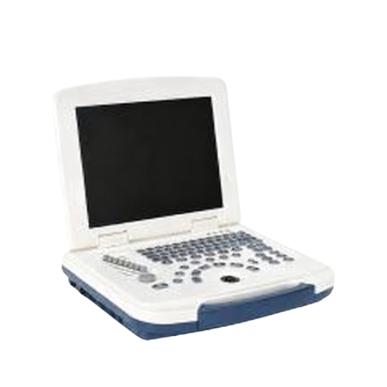
ArKang's AKB580 portable black and white ultrasound machine is a basic two-dimensional ultrasound machine that provides essential diagnostic functions for a range of applications, from obstetrics and gynecology to abdominal and small organ imaging.
3D ultrasound machines can scan tissue cross-sections from multiple angles, reconstructing data and generating three-dimensional images. Compared to two-dimensional ultrasound machines, 3D ultrasound machines overcome the planar limitations of 2D ultrasound, reconstructing the three-dimensional morphology of organs or lesions, making structural relationships more intuitive. They also allow for multi-plane rotation and magnification observation, avoiding missed diagnoses caused by the angle of the 2D ultrasound plane. Of course, 3D ultrasound machines offer faster examination speeds and shorter patient waiting times.
3D ultrasound is not only widely used in fetal, cardiac, obstetric, abdominal, and breast examinations, but it also provides far more intuitive three-dimensional images than 2D ultrasound, giving doctors crucial diagnostic insights and effectively improving the level of diagnosis and treatment.
4D ultrasound machines add a third dimension—time—to the three-dimensional model. A 4D ultrasound machine can accurately capture the morphology of organs/tissues in motion, offering strong visual clarity and real-time observation, significantly improving diagnostic efficiency in complex scenarios.
4D ultrasound machines offer high imaging resolution, are safe and radiation-free, and can clearly display minute structures. Besides applications in obstetric fetal examinations, cardiac disease examinations, and abdominal and organ examinations, they can also be used for real-time surgical monitoring, improving surgical precision and reducing intraoperative damage.
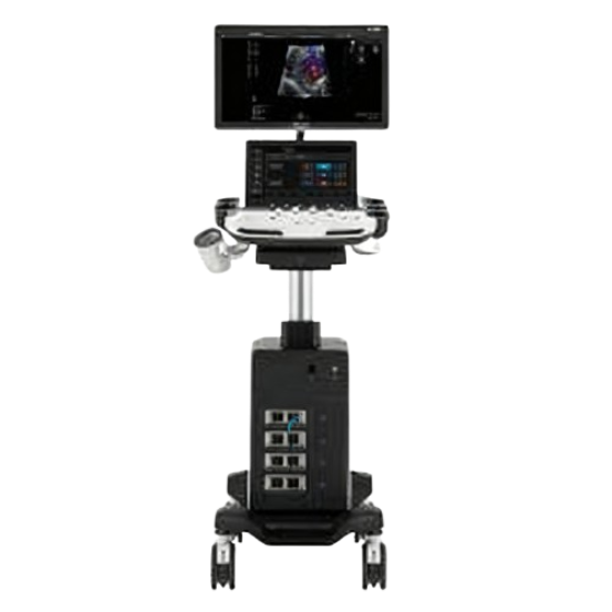
For example, ArKang's SonoScape P11 Elite model is a classic 3D/4D color Doppler sonography machine. It boasts advanced imaging capabilities, a competitive price, and excellent after-sales service, making it a popular choice among 3D/4D sonography machines.
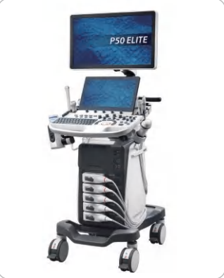
Doppler ultrasound utilizes the Doppler effect to measure blood flow and assess intravascular pressure in real time. The core feature of color Doppler ultrasound is its two-dimensional imaging combined with color blood flow display.
Its main principle is that the instrument emits ultrasound waves that penetrate the human body, receiving the reflected echoes from organs and tissues. These echoes are processed to form a two-dimensional cross-sectional image for observing the morphology and structure of organs. When ultrasound waves encounter moving red blood cells, the frequency of the reflected echo changes; this frequency shift is called Doppler shift. By detecting this Doppler shift, it is converted into color codes (typically red indicates blood flow towards the probe, and blue indicates flow away from the probe), which are then superimposed on the two-dimensional image to form a color blood flow map. By calculating the frequency shift data, parameters such as blood flow velocity, resistance index, and pulsatility index can be obtained, allowing for an objective assessment of hemodynamic status.
Besides its applications in obstetric examinations, abdominal examinations, and the diagnosis of heart diseases, color Doppler ultrasound is also helpful in diagnosing circulatory system diseases, arterial and venous diseases.
Sonoscape Doppler series is a popular brand, and ArKang, as a senior provider of Sonoscape Doppler ultrasound, has won the market's widespread trust with more than 23 years of professional experience.
The transesophageal echocardiography (TEE) probe is inserted through the esophagus, directly close to the back of the heart, avoiding the obstruction of ultrasound waves by lung air. It directly acquires detailed images of the heart structure from within the body, overcoming the blind spots of conventional transthoracic ultrasound.
The imaging resolution of TEE is significantly higher than that of transthoracic ultrasound, clearly displaying fine structures such as heart valves, atrioventricular septum, and coronary artery ostia. Simultaneously, it allows for real-time intraoperative monitoring without interfering with surgical procedures, meeting the dynamic assessment needs of cardiac surgery.
Specific applications of TEE include the diagnosis of complex heart diseases, intraoperative monitoring during cardiac surgery, assessment of coronary artery disease, and evaluation of thrombosis and cardiac function.
Endoscopic ultrasound (EUS) integrates endoscopy and ultrasound technologies. The endoscope guides the probe to the target site, while ultrasound provides deep structural imaging, combining "direct observation" with "penetrating detection."
EUS offers high imaging resolution, clearly displaying the layered structure of the digestive tract wall (mucosa, muscle layer, etc.). It can also penetrate the digestive tract wall to detect surrounding organs (such as the pancreas, bile ducts, and spleen), addressing deep lesions that conventional endoscopy cannot reach.
Endoscopic ultrasound can be applied to the diagnosis of digestive tract diseases, examination of surrounding organs, and detection of minute lesions.
Portable ultrasound machines are popular in recent years due to their portability and compact size, allowing for examinations anytime, anywhere. While the imaging quality of a portable ultrasound machine may not match that of a high-end cart-mounted machine, it is perfectly adequate for basic diagnostics in field settings and emergency rooms, helping healthcare professionals make quick decisions.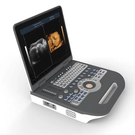
For example, ArKang's AK-UTS1600 is a classic portable color Doppler ultrasound system, small and lightweight at only 6.5 kg. This system is ideal for clinicians who need reliable, high-quality imaging in various environments, from hospital departments to mobile healthcare services.
In the musculoskeletal field, sonogram machines are primarily used for diagnosis, assessment, and rehabilitation. These ultrasound machines visualize the morphology and structure of the musculoskeletal system, allowing for the identification of muscle tears, ligament injuries, and assessments of joint effusions, cartilage damage, and space-occupying lesions in the soft tissues surrounding bones.
Color Doppler ultrasound is a commonly used tool for musculoskeletal diagnosis, primarily used for morphological observation and blood flow assessment of muscles, tendons, ligaments, and joints.
Three-dimensional/thinking sonogram machines are suitable for the three-dimensional assessment of complex muscle injuries (such as articular cartilage damage and tendon insertion lesions), offering high diagnostic accuracy.
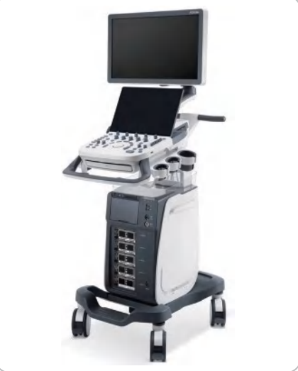
For example, ArKang's SonoScape P20 Elite trolley-mounted 3D/4D color Doppler ultrasound system excels in musculoskeletal applications, clearly visualizing muscle morphology and helping clinicians accurately diagnose muscle injuries and other conditions.
In gynecology, sonogram machines are primarily used for the diagnosis of gynecological diseases and pregnancy monitoring in women. Gynecological examinations mainly focus on examining female pelvic organs, such as the size of the uterus, ovaries, and fallopian tubes. The most basic two-dimensional ultrasound machine can be used for pregnancy monitoring, is radiation-free, and easy to operate.
B-mode ultrasound (two-dimensional ultrasound) can be used for routine gynecological screening, suitable for basic morphological examinations of the uterus and ovaries, confirmation of early pregnancy gestational sacs, etc. It is fast and cost-effective.
Color Doppler ultrasound can be used for precise diagnosis of gynecological diseases, assessing tumor blood supply through blood flow signals (e.g., malignant tumors often have rich blood flow), monitoring placental blood flow, etc. However, the functions of two-dimensional ultrasound are generally sufficient for monitoring throughout all stages of pregnancy.
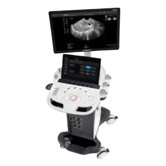
For example, the ArKang SonoScape P9 Elite trolley-type color Doppler ultrasound system is very suitable for obstetrics, gynecology, follicle monitoring, and fetal malformation screening. Of course, some basic two-dimensional ultrasound machines, such as the AKB-K10, can be used for all stages of prenatal care.
In urology, sonogram machines can examine organs such as the kidneys, ureters and bladder, prostate, and male reproductive organs for defects. They can also detect urinary tract infections and urination problems.
B-mode ultrasound is a basic tool for routine screening in urology, while color Doppler ultrasound and 3D/4D sonogram machines are more suitable for precise diagnosis and assessment of complex conditions in urology.
For example, ArKang's SonoScape P11 Elite trolley-type 3D/4D color Doppler ultrasound system features 2D/3D/4D imaging modes, providing clear images and applicable to general imaging, obstetrics and gynecology, vascular surgery, urology, cardiology, and other fields.
A sonography machine is an ideal tool for examining the abdomen, using ultrasound to diagnose diseases of the liver, gallbladder, pancreas, spleen, and gastrointestinal tract.
Two-dimensional ultrasound is a basic screening tool for abdominal diagnosis. Color Doppler ultrasound is used for precise diagnosis, assessing organ blood supply (e.g., blood supply characteristics of liver cancer) and hepatic artery/portal vein blood flow status through blood flow signals, and assisting in differentiating between benign and malignant tumors. Three-dimensional/thinking ultrasound is suitable for three-dimensional assessment of complex lesions.
Using standard ultrasound and Doppler ultrasound to generate echocardiograms, it is possible to observe the size of the atria and ventricles, screen for congenital heart disease, and quantitatively assess cardiac function, evaluating the heart's systolic/diastolic capacity and determining the type and severity of heart failure. Ultrasound examination allows patients to detect heart problems promptly, making it one of the most crucial treatment methods for patients with heart failure.
When women undergo mammography, the use of an X-ray machine inevitably exposes them to some radiation, especially during pregnancy, which can easily damage the image of both the woman and the fetus. Ultrasound examinations, however, do not pose this risk. Currently, breast ultrasound is increasingly becoming the primary choice for women's breast examinations due to its safety and convenience. Basic two-dimensional ultrasound can usually clearly display the structure of breast tissue and the morphology of nodules, suitable for initial screening and regular checkups. For differentiating between benign and malignant tumors, color Doppler ultrasound is more suitable.
Many animals are also suitable for ultrasound examination, such as in animal reproduction and obstetrics. This is the most common scenario. Veterinarians can easily determine the number of fetuses and monitor fetal development throughout the gestation period using ultrasound.
Ultrasound itself does not cause any harm to animals and does not subject them to the stress of traditional surgery. Therefore, ultrasound is widely used and popular in animal examinations. Whether in veterinary hospitals, farms, or even some domestic pets, veterinary ultrasound machines are often provided by veterinarians.
ArKang Company in Foshan is a professional medical equipment supplier, and also provides veterinary ultrasound machines. For 23 years, ArKang Company has won the trust of its customers with competitive prices and excellent product quality.
Philips is a well-known ultrasound machine brand. Its ultrasound equipment excels in user-friendliness, with many models equipped with touchscreens for simple and intuitive operation. Its ClearVue series is particularly popular in obstetric ultrasound due to its high image clarity. However, it may not be as advanced as GE Healthcare in some high-end features.
Top-of-the-line models such as the SC2000 and the professional mammography S2000 ABVS offer exceptionally clear image quality with excellent detail processing. They also feature unique vector analysis technology and distinctive elasticity imaging capabilities.
However, Siemens software is somewhat cumbersome, the system can be complex, boots up slowly, and sometimes crashes. Their market strategy is relatively conservative.
Mindray Medical is a leading Chinese medical device manufacturer. Its ultrasound machines offer high cost-performance and continuously improving product functionality. For example, Mindray's new generation whole-body application-oriented economical color Doppler ultrasound, the DC-30, features broadband frequency-shift harmonic imaging and intelligent speckle noise suppression imaging technologies, effectively reducing noise and improving image resolution. It also comes equipped with iScanHelper built-in ultrasound teaching software to help users quickly learn ultrasound scanning.
However, Mindray Medical currently lags slightly behind other internationally renowned brands in the high-end imaging market.
GE Healthcare's ultrasound machines are renowned for their excellent image quality and stability, suitable for various clinical scenarios. Its high-end models, such as the Logiq E9, feature advanced image processing technology, facilitating real-time imaging and storage, and are widely used in cardiovascular and obstetric/gynecological examinations.
However, GE Healthcare currently has a long product line, its latest ultrasound machines lack innovation, and some of its high-end ultrasound machines are relatively expensive.
Foshan ArKang Medical is committed to providing high-quality medical equipment at highly competitive prices. Its product line covers portable ultrasound machines, handheld ultrasound machines, and trolley-type color Doppler ultrasound systems, and is widely recognized in the market. Although its brand awareness is not yet as high as industry giants, ArKang offers an ideal choice for customers seeking high cost-performance, thanks to its clear image quality and reasonable pricing.—— Learn More
By considering some important factors related to ultrasound, you can control your budget and purchase the right ultrasound machine for your needs at the most affordable price.
Basic diagnostic screening ultrasound is sufficient, but for more precise diagnosis, a 3D/4D ultrasound machine is needed. Determining the direction of blood flow requires a color Doppler ultrasound system. First, clearly define your diagnostic needs, then purchase a suitable ultrasound machine. Of course, the clearer the ultrasound image, the better for your diagnostic findings.
Different ultrasound machines may be suitable for examining different areas. Determining your clinical application needs is the primary prerequisite for purchasing an ultrasound machine. For example, cardiology departments may require a 3D/4D ultrasound machine for accurate diagnosis, while assessing blood flow velocity at the heart valve orifices requires a Doppler ultrasound system.
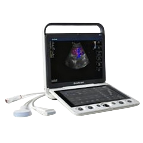
Currently, many ultrasound machines are applicable to a wide range of fields. A single ultrasound machine can be used in gynecology, urology, abdominal surgery, cardiology, etc. The ArKang SonoScape S9 portable color Doppler ultrasound system is particularly recommended. This system combines portability and high performance, demonstrating excellent versatility in multiple disciplines, including cardiology, radiology, abdominal surgery, obstetrics and gynecology, and urology. It won the 2014 iF Design Award, and its reasonable price makes it an ideal choice for many medical institutions.
When purchasing an ultrasound machine, remember to specifically inquire with the supplier about the machine's application areas and special functions to help find the right ultrasound equipment for your needs.
There are various types of probes, such as convex array probes, linear array probes, and phased array probes. Different types of probes are suitable for different examination sites and clinical needs. At the same time, the frequency range of the probe is also important. High-frequency probes have high resolution but low penetration, while low-frequency probes have the opposite. The appropriate frequency probe should be selected based on the actual examination requirements.
When purchasing ultrasound equipment, probes are a key factor to consider. First, be sure to confirm whether the supplier's quotation includes probes. Second, understand the number of probes supported by the main unit and their interchangeability, which directly affects the equipment's flexibility in handling different clinical examinations. A reasonable probe configuration is key to controlling procurement costs and improving future efficiency.
A simple and intuitive user interface allows medical staff to quickly become familiar with the equipment, reducing operation time and errors, and improving work efficiency. It's also important to inquire with the supplier about comprehensive training services. During negotiations, we need to secure more of our rights.
Choosing a trustworthy brand when purchasing ultrasound equipment can save you a lot of trouble, as well-known brands usually offer higher product quality and comprehensive after-sales service. ArKang Medical has been exporting medical equipment for over 23 years, and its products are known for their stability and reliability. Furthermore, it has a mature after-sales service system, reducing unnecessary hassles for customers.
When purchasing ultrasound equipment, we shouldn't solely pursue high performance. We also need to consider our budget. Therefore, when purchasing ultrasound equipment, we must comprehensively consider factors such as the equipment's performance, functions, and quality. We also need to consider the cost of probe replacement, energy consumption, maintenance costs, and other subsequent operating expenses. It's important to know that some ultrasound machines may have low initial purchase prices but high operating costs, which may not be cost-effective in the long run.
Currently, ultrasound machines have become one of the best choices for medical diagnosis due to their safe, radiation-free, and harmless characteristics. There are various types of ultrasound machines on the market, and different types of sonograph machines may be suitable for different fields. Understanding the uses and applications of ultrasound machines will be of great help in our procurement.
Ultrasound machines mainly include 2D ultrasound, 3D ultrasound, 4D ultrasound, color Doppler ultrasound, endoscopic ultrasound, and portable ultrasound machines.
Ultrasound equipment refers to devices that utilize sound waves at frequencies above the audible range to perform various functions, including generating large amounts of sound energy, surface cleaning, and acoustic manipulation of particles.
Ultrasound imaging (sonography) uses high-frequency sound waves to view inside the body. Because ultrasound images are captured in real-time, they can also show movement of the body's internal organs as well as blood flowing through the blood vessels.
Ultrasound machines can be used in various fields such as abdominal, cardiac, obstetric, gynecological, urological, and cerebrovascular examinations; breast examinations; small tissue sample examinations; and pediatric examinations. Different fields may require different ultrasound machines.
Well-known brands include Philips, Siemens, Mindray Medical, and GE Healthcare, but their ultrasound machines are relatively expensive. Alternatively, you could choose the emerging brand ArKang, which offers more competitive pricing.
The answer depends entirely on the primary examinations they will perform (e.g., abdominal, vascular, cardiac, MSK, OB/GYN).
You need to confirm whether the machine is optimized for your workflow, whether you work in radiology, cardiology, emergency medicine, point-of-care ultrasound (POCUS), veterinary medicine, etc.
The total price of an ultrasound machine typically includes not only the ultrasound machine itself but may also include the cost of the required probes, software licenses, and installation.
There are usually several financing options available, including lease-purchase agreements, loans, or subscription models, which you can choose based on your financial situation and operational goals.
After purchasing ultrasonic equipment, ongoing costs for consumables such as ultrasonic gel, probe sleeves, and printing paper must be considered for its operation. Additionally, please ensure that software upgrades and annual service contracts are included in the total equipment price. Furthermore, it is recommended to prioritize manufacturers that offer comprehensive warranty services, as this will effectively reduce long-term repair costs due to unexpected malfunctions.


